Echocardiography
Echocardiography is the principal investigation that allows the diagnosis of mitral regurgitation or stenosis and heart function. It uses ultrasound waves to image heart structures, and to observe and quantify the blood flow through heart valves and chambers.
Transthoracic echocardiogram (TTE) is the most commonly performed examination. During the examination, you will lie down on a couch. The test is performed by using gel on the skin and a device that is attached to the ultrasound machine (transducer). The transducer is located firmly against your skin to acquire images of the heart and valves.
Transoesophageal echocardiogram (TOE): In some patients, we may need to use a different probe that is inserted down the oesophagus. This will allow for a more accurate assessment of the mitral valve morphology and function. The technique is also used to guide surgical and transcatheter mitral procedures.
A stress echocardiogram may be performed in some patients to observe heart function during exercise. This will be performed as a transthoracic echocardiogram whilst you are exercising (eg. on a bike or treadmill). The test helps us to understand the function of the heart chambers and valves during exertion.


Computed Tomography (CT scan)
Our expert team provides the following services:
- CT Coronary artery calcium scoring and angiography
- CT for planning Transcatheter mitral and aortic valve implantation (TMVI, TAVI)
- CT for planning minimal access/endoscopic mitral and aortic valve surgical procedures
What is a CT Scan?
A CT scanner uses X-rays to produce images of the body organs. The
images are used to guide mitral valve treatment and plan the procedure
safely.
When having a CT scan, you lie on a couch that moves slowly into the
scanner. Only part of your body will be inside the machine. The scan only
takes few seconds and you may be asked to hold your breath during the
scan.
We may need to inject contrast agent (dye) into your veins during the
scan to allow better visualisation of your heart and vessels. We
recommend that you come well hydrated prior to your scan.
How does CT scan results help patients undergoing mitral valve procedures?
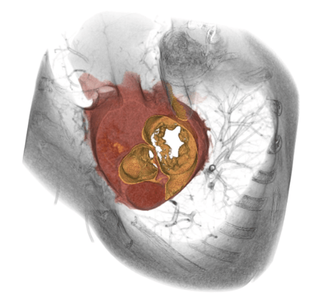
CT scan images are used to perform a comprehensive assessment prior to transcatheter or surgical mitral valve procedures. A cardiac radiologist uses advanced software to see the inside of your body. The processed images help cardiologists and cardiac surgeons to plan the most appropriate and safest procedure for each individual patient.
For example, we look at the size, and the distribution of the calcification around the mitral valve prior to a procedure. A CT scan is also used to show the safest access for the procedure.
In patients with higher cardiovascular risk, a CT coronary angiogram is used to exclude coronary artery plaque disease and stenosis.
Our Team